
The Pelvis and Hips - Ligaments
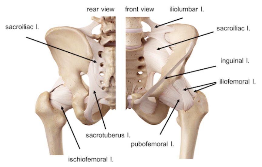
Sacroiliac ligaments
-
This ligament is better considered as a thickening of the anterior aspect of the sacroiliac joint capsule. It connects the anterior surface of the lateral sacrum to the auricular margin of the ilium. It is also considerably thinner than the posterior sacroiliac ligament.
-
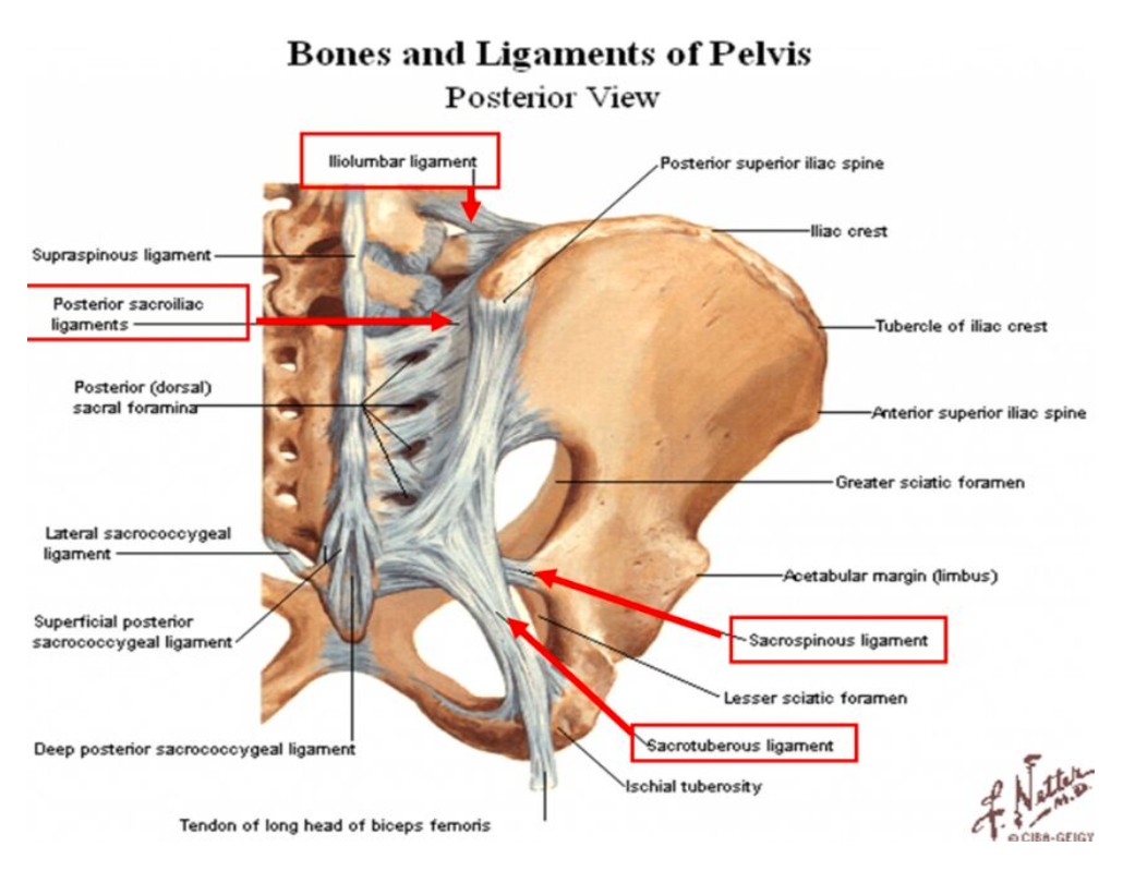
This can be further divided into the short (deep) and long (superficial) components. The deep/short fibres run almost horizontally. They connect the first and second transverse tubercles, that arise from the posterior surface of the sacrum, to the iliac tubercle. The superficial/long fibres of the posterior sacroiliac ligament run in an oblique vertical direction. They attach the third and fourth transverse tubercles, that arise from the posterior surface of the sacrum, to the posterior superior iliac spine.
-
The dorsal interosseus ligaments are a series of short and strong ligaments that run in a nearly horizontal direction to prevent subluxation or dislocation of the sacroiliac joint.
Other ligaments of the sacroiliac joint
-
This ligament extends from the lateral surface of the transverse process of the fifth lumbar vertebrae, and inserts onto the inner lip of the iliac crest. It stabilizes the joint superiorly and strengthens the joint overall.
-
This is a triangular-shaped flat ligament that runs from the lower transverse tubercles of the sacrum and the superior part of the coccyx, to the ischial tuberosity. The broad base of the ligament arises from the posterior superior iliac spine, as well as the posterior sacroiliac ligaments. The fibres run in an oblique direction, and converge to a narrow point of fibres that insert onto the medial surface of the ischial tuberosity. The ligament is pierced by the coccygeal branches of the inferior gluteal nerve, as well as cutaneous branches of the coccygeal plexus. The most inferior part of the gluteus maximus muscle also attaches to the sacrotuberous ligament, as do some of the fibres of biceps femoris. The sacrotuberous ligament runs behind the sacrospinous ligament along its entire course.
-
This is another thin triangular-shaped ligament of the pelvis. It arises from the lateral margin of the sacrum and the coccyx, and inserts onto the ischial spine. The fibres do mingle and merge with the sacrotuberous ligament. The sacrospinous ligament covers the coccygeus muscle along its entire length. The greater sciatic notch forms above the ligament, and the lesser sciatic notch forms below the ligament. The presence of the ligament divides the notches into greater and lesser sciatic foramina.
The sacrococcygeal symphysis is supported by five ligaments:
Anterior sacrococcygeal ligament – a continuation of the anterior longitudinal ligament of the spine, and so connects the anterior aspects of the vertebral bodies.
Deep posterior sacrococcygeal ligament – connects the posterior side of the 5th sacral body to the dorsal surface of the coccyx.
Superficial posterior sacrococcygeal ligament – attaches the median sacral crest to the dorsal surface of the coccyx.
Lateral sacrococcygeal ligaments – run from the lateral aspect of the sacrum to the transverse processes of Co1.
Interarticular ligaments – stretch from the cornua of the sacrum to the cornua of the coccyx.
Ligaments of the pubic symphysis:
The superior pubic ligament runs from the left to right pubic crest, attaching to the pubic tubercles. This is reinforced by the tendons of the rectus abdominis, external oblique, gracilis and hip muscles
The posterior pubic ligament extends from the left to right inferior pubic ramus (aka arcuate ligament), an forms the upper boundary of the pubic arch
These two ligaments are significantly stronger than the other two
The anterior pubic ligament consists of several superimposed layers, which pass across the front of the articulation.
The superficial fibres pass obliquely from one bone to the other, decussating and forming an interlacement with the fibres of the aponeuroses of the Obliqui externi and the medial tendons of origin of the Recti abdominis
The deep fibres pass transversely across the symphysis, and are blended with the fibrocartilaginous lamina.
The posterior pubic ligament consists of a few thin, scattered fibers, which unite the two pubic bones posteriorly.
Other ligaments of the hips & pelvis:
The lumbosacral ligaments are found bilaterally, connecting the inferior aspect of the transverse processes of L5 to the lateral surfaces of the sacrum
The inguinal ligament is the lower border of the aponeurosis of the Obliquus externus, and extends from the anterior superior iliac spine to the pubic tubercle. From this latter point it is reflected backward and lateral to be attached to the pectineal line for about 1.25 cm., forming the lacunar ligament.
The lacunar ligament is that part of the aponeurosis of the Obliquus externus which is reflected backward and lateralward, and is attached to the pectineal line.
The pectineal ligament (sometimes known as the inguinal ligament of Cooper) after Astley Cooper) is an extension of the lacunar ligament that runs on the pectineal line of the pubic bone. The pectineal ligament is the posterior border of the femoral ring.